Scanning Electron Microscope (SEM) Lab

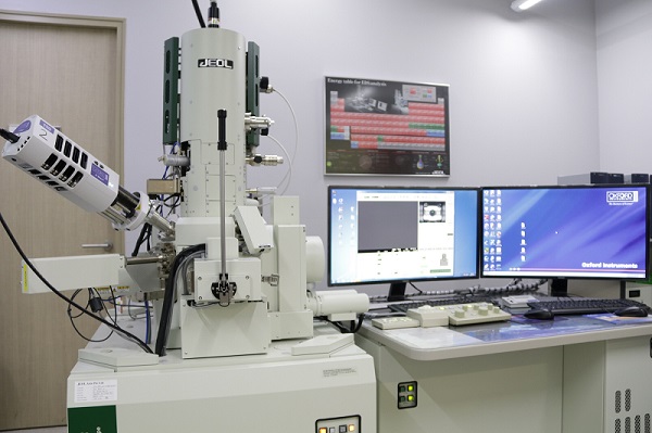
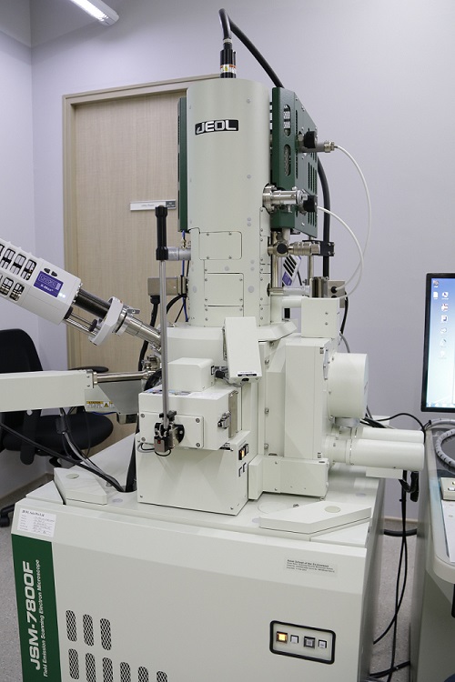
The Scanning Electron Microscope (SEM) is used for imaging minerals and other geological, biological, and environmental materials at the micrometer scale. Our JEOL JSM-7800F Field Emission SEM is equipped with secondary electron (SE) and backscattered electron (BSE) detectors and can operate in either high vacuum mode or low vacuum (variable pressure) mode. The pressure range over which the system can operate makes it possible to image nonconductive materials with or without coating.
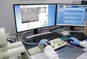
Two Oxford X-Max 150 mm2 energy dispersive x-ray spectrometers (EDS) are used to qualitatively measure major element abundances in minerals, glasses, and other materials. The EDS detectors are also used for making qualitative element profiles (line scans) and maps of minerals. Crystal structures and orientations can be identified using the Oxford NordlysNano electron back scatter diffraction (EBSD) detector. Oxford’s AZtec microanalysis software is used to run the EDS and EBSD detectors. The software is also used to quantify EDS analyses/maps using standard reference materials and automatically mosaic high-resolution electron images and EDS maps from large (up to several cm) areas.
A Leica EM ACE600 high-vacuum coater is used to apply a thin film of carbon or metal (e.g. gold, iridium) onto the surface of nonconductive samples prior to imaging and analyses. This completely automated instrument is used for metal sputtering, carbon thread evaporation, carbon rod evaporation, E-beam evaporation, and glow discharge, providing the perfect coating for many different applications. A Cressington 208Carbon coater is also housed in the SEM lab.
Common examples of applications include:
- Identifying minerals and their chemical compositions from all rock types.
- Making BSE images of entire thin sections used to investigate textural complexities of mineral, glass, and vesicle populations and to identify areas that will be studied further.
- Making BSE and compositional maps of complexly-zoned phenocrysts in volcanic rocks to qualitatively understand the thermal evolution of the crystals.
- Obtaining compositional profiles across volcanic crystals to be used for diffusion time scale modelling.
- Investigating morphologies of quartz sand grains to distinguish different sediment sources.
- Differentiating calcite from aragonite in modern and ancient carbonates.

